The following is my attempt to explain the working of the brain by looking at the different sections of the brain and then by looking more closely at a microscopic level of the brain. I have kept scientific terms to a minimum; choosing instead to use analogies wherever I can to describe both appearance and function. Where I feel it is useful I have chosen terms about the brain that have been embedded in our common language, rather than the more technical names assigned by the science that is neurology. I use only single words to name individual parts of the brain despite many of these having more than one name. Hence, I use ‘brain cell’ instead of ‘nerve cell’ or ‘neuron’ and ‘hind brain’ instead of the ‘archipallium’ or ‘reptillian brain’. I have written the following simplified description to help in establishing my own understanding of the brain. I have no idea how useful this will be to others that read it. I would suggest that, for those of you that wish to truly understand more about the brain, you do your own reading about the brain, found in texts that take up the topic in great detail.
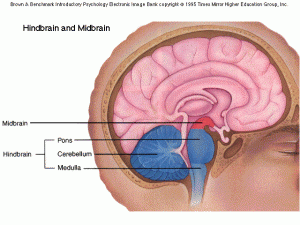
Here it is, as simple as I can make it. The brain has 4 main parts to it. The hind brain located at the back of the head, the mid brain, located where its name sake suggests, the crown which is the cover of the brain that gives it its walnut appearance, and the bridge connecting the two halves of the crown.The hind brain is the most active area of the brain and is responsible for our balance, coordination and controlled movements. “Hardwired attitudes, emotional reactions, repeated actions, habits, conditioned behaviours, unconscious reflexes, and skills that we have mastered are all connected to and memorized in the hind brain.” – (Dispenza, J. (2007) Evolve Your Brain. Health Communications Inc, p110.
The midbrain is about the size of an apricot. It controls our body temperature, blood sugar levels, blood pressure, digestion, hormone levels etc. All signals that the body receives come through the midbrain which connects it to the correct part in the crown. The midbrain produces the chemicals necessary to monitor the body as well as the chemicals that make us feel the way we think. In dangerous situations it gets us ready by preparing our bodies to run or fight. It holds our internal clock and chemically controls our patterns of sleep. The mid brain stores the kind of long term memories that come from associative learning. It also stores the strong emotions of aggression, joy, sadness and fear.
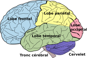
The crown is where our thinking, learning and most of our remembering take place. It comes in two layers. The outer wrinkled layer is 3 to 5 mm thick and is often referred to as our grey matter. The crown is divided into a left and right side which is connected by the bridge which gives the crown the ability to observe the world from two different points of view. Each side of the crown is divided into 4 lobes (areas). Each lobe processes different sensory information, motor abilities, and mental functions and is assigned to perform different tasks. The front lobe is where we focus our attention and coordinate nearly all functions of the rest of the brain. This is the area of the brain where we focus on our desires, create ideas and make conscious decisions. It is what gives us our free will. No wonder I am always clutching it with my hand.
The lobe back from the frontal lobe is the parietal lobe which processes our sensations related to touch as well as co-ordinating some language functions. The temporal lobes are located on the left and right side of the brain and are largely concerned with processing sounds, perception, learning, language, smell and memory. Finally, the occidental lobe is located at the back of the brain and deals with processing data from the outside world in order for us to see.
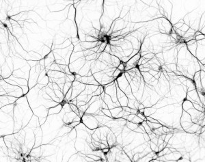
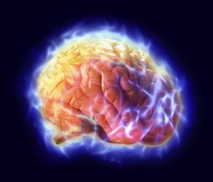
We all have a similar image of the brain in our brains, if you know what I mean. That picture of a large wall nut that has been moulded to fit inside the shape of our skull. A piece of human anatomy that can be held in the hands, sliced and diced. This visual image that we hold along with my description of the different sections of the brain helps create an overall picture of a solid structure organised into clear sections, with each section solely responsible for performing certain tasks. Unfortunately, this kind of image misrepresents how the brain actually operates. It is far better to think of the brain as an electrically charged bowl of billions of microscopic pieces of cotton; each piece of cotton resembling a leafless oak tree. These billions of oak trees with their web of branches and roots never actually touching each other but separated by the smallest of gaps. These cotton oak trees represent brain cells; their roots are capable of receiving chemical shots that jump from the ends of the branches of other oak trees. These chemical shots can either fire up an electrical impulse in the receiving tree which may then shoot up the tree until it reaches the end of a branch causing another chemical reaction and a repeat of the cycle, or they can help to put an immediate end to the process.
Our brain cells are organised in a way that facilitates the setting up of paths that actually give us our individual uniqueness. The chemicals that are produced in brain cells by our very thoughts is what determines how we feel. It is our nervous system that connects the environment to the body, the body to the brain, and the brain to the body.

This oversimplification of the how the brain works doesn’t offer much to a teacher looking for ways to improve his lessons, but it hopefully sets up a picture of the brain that will help when we begin to consider how the brain actually learns. My guess is that it is the process of trying to simplify the way the brain works that is far more useful than the end result of this article. I found it a valuable exercise and would encourage other brain novices to do the same.


